Back to blog

Coauthored with Patrick McDaniel from HTL
From dermal fillers to eye drops and from drug delivery to tissue engineering, hyaluronic acid’s versatility has been fueling this polysaccharide’s surging popularity in recent years. Despite all its desirable attributes – biocompatibility, ease of functionalization and biodegradability to name a few - limitations of HA remain. One such limitation is the fact that while viscous, HA is not rigid and would collapse under its own weight if any kind of three-dimensional shape was to be formed. Another limitation is the relatively fast degradation time in-vivo. There are certainly applications where fast degradation is an asset, but for any kind of sustained-release or viscosupplementation application, a slower degradation time is the difference between the HA being efficacious or not.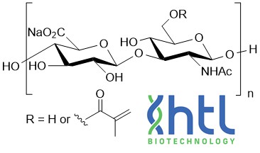
One straightforward solution to overcoming the limitations caused by the HA’s fluidity and rapid degradation in vivo is to modify the hyaluronic acid with methacrylate. Methacrylated HA (HA-MA) allows the HA to be polymerized by free radicals via the addition of a photosensitive initiator. The resulting photocured, crosslinked hydrogel will have increased rigidity and be more resistant to degradation, even at low concentrations of 10% or less. With the enhanced rigidity, researchers can now bioprint, or otherwise fabricate, 3D structures and crosslink them with UV light immediately after gel deposition to maintain the shape. This makes HA-based scaffolds, organoids, cell culturing and disease models possible. The slower degradation time of the HA-MA hydrogels and structures also allows for sustained release drug delivery systems to be designed based on the degradation profile and the dosage of the drug vs. time. One bonus feature of the methacrylated HA gels is that everything from genes to cells to whole organisms can be encapsulated and protected from harsh physiological conditions in vivo.
When crosslinking via photopolymerization, the starting molecular weight of the HA should be optimized – less than 1000kDa is most common - but the dominant factor in determining the physical properties is the degree of substitution (DS). The higher the DS, the more vinyl conversions which leads to a higher degree of polymerization which means lower swelling ratios, slower degradation times and higher hydrogel shear moduli along with other rheological measurements. It’s worth noting, considering how the most popular use for methacrylated HA is to make 3D structures for live cells, that the DS of methacrylated HA does not affect cell attachment and proliferation.
The degree of substitution can range from low (<30%) to high (>60%). Below is a table of example methacrylated HA applications and the concentrations, molecular weight and DS:
| Application | HA MW | DS | Concentration |
|---|---|---|---|
| Scaffold for Intervertebral Disc Repair | 400kDa | 25% | 1% |
| Hydrogel Scaffold for Tumor Modeling | 300kDa | 20-60% | 10% |
| Hydrogel for Biologic Encapsulation | 700kDa | 94% | 3% |
| Hydrogel for Wound Healing Dressing | 70kDa | N/A | 4% |
| Carrier for Stem Cell Delivery | 400kDa | 50% | 1.5% |
In conclusion, methacrylated hyaluronic acid hydrogels offer a versatile platform for various in vitro applications. Their ability to mimic the natural microenvironment of tissues, coupled with their tunable properties, makes them valuable tools for tissue engineering, drug delivery, and stem cell research. As researchers continue to refine and expand their understanding of HA-MA hydrogels, their potential for advancing regenerative medicine and improving patient care becomes increasingly evident.
Methacrylated Hyaluronate 100 kDa
Methacrylated Hyaluronate 250 kDa
Methacrylated Hyaluronate 850 kDa
References
- Wang, J. et al, (2022) Photo-crosslinked hyaluronic acid hydrogel as a biomimic extracellular matrix to recapitulate in vivo features of breast cancer cells. Colloids and Surfaces B: Biointerfaces 209, 112159
- Huang, L. et al, (2022) ROS-responsive hyaluronic acid hydrogel for targeted delivery of probiotics to relieve colitis. International Journal of Biological Macromolecules 222, 1476–1486
- Wang, D. et al, (2022) Hyaluronic acid methacrylate/pancreatic extracellular matrix as a potential 3D printing bioink for constructing islet organoids. Acta Biomaterialia, 4:56
- Chang, L. et al, (2023) Hyaluronic acid methacrylate/laponite hydrogel loaded with BMP4 and maintaining its bioactivity for scar-free wound healing. Regenerative Biomaterials, 10, rbad023
- Oulong, L. et al, (2020) Expanding and optimizing 3D bioprinting capabilities using complementary network bioinks. Applied Sciences and Engineering
- Gong, M. et al, (2022) A dopamine-methacrylated hyaluronic acid hydrogel as an effective carrier for stem cells in skin regeneration therapy. Cell Death & Disease, 13, Article number: 738
- Chen, P et al, (2017) Photo‐crosslinked gelatin‐hyaluronic acid methacrylate hydrogel‐committed nucleus pulposus‐like differentiation of adipose stromal cells for intervertebral disc repair. Journal of Tissue Eng Regen Med; 13:682–693.
- Bencherif, S. et al. (2008) Influence of the degree of methacrylation on hyaluronic acid hydrogels properties. Journal of Biomaterials; 11:47
0.2
/ 0.3
Related Articles
Stay informed with our informative blog posts.
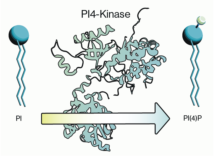
Why you should care about PI4-Kinase activity
PI4-kinase, or phosphatidylinositol 4-kinase (PI4K), is an enzyme involved in the synthesis of phosphatidylinositol 4-phosphate (P
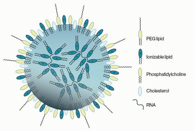
Lipid Nanoparticles for RNA Delivery
Genetic therapies have now been pursued for several decades and close to a dozen DNA-based gene therapies have gained regulatory a
Semi-quantitative PEth ELISA holds promise for simplifying clinical analysis of alcohol consumption
Coauthored with Jeff Johnson and Melissa Burback According to a May 2022 report from the World Health Organization (WHO), 3 millio
Methacrylated Hyaluronic Acid: Benefits and Applications
Coauthored with Patrick McDaniel from HTL From dermal fillers to eye drops and from drug delivery to tissue engineering, hyaluroni
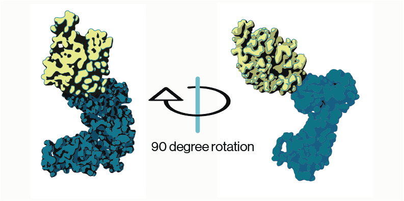
PIKfyve: function and inhibition as therapeutic opportunities
PIKfyve, also known as PIP5K3, is a lipid kinase that synthesizes two rare phosphoinositides, PI(5)P and PI(3,5)P2 (Figure 1). The
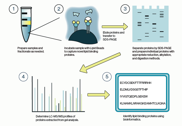
Identifying lipid-protein interactions with Lipid Beads and LC/MS
Lipid Coated Beads are lipid-protein interaction tools originally conceived around eight phosphatidylinositol phosphate coatings t
0.3
/ 0.3
Get in Touch
If you have any questions or would like to learn more about our services, feel free to reach out to us. We’re here to help!
Biosciences